


 12:30 PM
 md arman

Bacteriology – Science that deals with Bacteria.
 This cellular compartment is found only in those bacteria that have both an outer membrane and plasma membrane (e.g. Gram negative bacteria). In the space are enzymes and other proteins that help digest and move nutrients into the cell.
 This is a lipid bilayer much like the cytoplasmic (plasma) membrane of other cells. There are numerous proteins moving within or upon this layer that are primarily responsible for transport of ions, nutrients and waste across the membrane.

b) Bacillus (Cylindrical) – Rod-Shaped. e.g. E. coli.

c) SPirillum (Spirocheate) – Spiral-shaped. e.g Treponema pallidum.

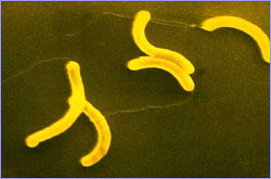
d) Vibrio – Comma-shaped. e.g. Vibrio cholerae
e) Filamentous – threadlike. e.g. Actinomyces israelli.

Bacteriology - A unicellular organisms without a true nucleus.
Mycoplasmas are bacteria that have no cell wall and therefore have no definite shape.
Internal Structure
Bacteria have a very simple internal structure, and no membrane-bound organelles.
Nucleoid: DNA in the bacterial cell is generally confined to this central region. Though it isn’t bounded by a membrane, it is visibly distinct (by transmission microscopy) from the rest of the cell interior.
DNA- Deoxyribo Nucleic Acid is single long circular molecule. It contains all genetic information for the structure & function of a bacterium under its optimum conditions.
Ribosome
Ribosome gives the cytoplasm of bacteria a granular appearance in electron micrographs. It is important in translating the genetic massage in messenger RNA ( Ribo Nucleic Acid) into the production of peptide sequences (proteins). It is responsible for protein synthesis.
Storage Granules
Nutrients and reserves may be stored in the cytoplasm in the form of glycogen, lipids, polyphosphate, or in some cases, sulfur or nitrogen.
Endospore
Some bacteria, like clostridium botulinum, form spores that are highly resistant to drought, high temperature and other environmental hazards. Once the hazard is removed, the spore germinates to create a new population.
Capsule
This layer of polysaccharide (sometimes proteins) protects the bacterial cell and is often associated with pathogenic vacteria because it serves as a barrier against phagocytosis by white blood cells.
Outer membrane
This lipid bilayer is found in Gram negative bacteria and is the source of lipo-polysaccharides (LPS) in these bacteria. LPS is toxic and turns on the immune system of, but not in Gram positive bacteria.
Cell wall
This is the complex & semi-rigid outermost layer. It gives shape to the bacterial cell & protects it from external environment. It is composed of peptidoglycan (polysaccharides + protein); the cell wall maintains the overall shape of a bacterial cell.
Periplasmic Space
This cellular compartment is found only in those bacteria that have both an outer membrane and plasma membrane (e.g. Gram negative bacteria). In the space are enzymes and other proteins that help digest and move nutrients into the cell.
Plasma membrane
 This is a lipid bilayer much like the cytoplasmic (plasma) membrane of other cells. There are numerous proteins moving within or upon this layer that are primarily responsible for transport of ions, nutrients and waste across the membrane.
Appendages:
Bacteria may gave the following appendages ( a tail or a limb attached to a major part):
Pili
Pili: These are hollow, hair like structures made of protein allow bacteria to attach to other cells. A specialized pilus, the sex pilus, allows the transfer from one bacterial cell to another. pili (sing; pilus) are also called fimbriae (sing; fimbria).
Flagella
The purpose of flagella (sing; flagellum) is motility. flagella are long appendages which rotate by means of a “motor” located just under the cytoplasmic membrane. Bacteria may have one, a few or many flagella in different positions on the cell.
Classification of Bacteria
Bacteria can be classified in many ways but three are important –
1. Classification on the basis of morphology (Shape & Size):
a) Coccus (Spherical) – round shaped. e.g. Streptococcus, Pneumococcus.

b) Bacillus (Cylindrical) – Rod-Shaped. e.g. E. coli.

c) SPirillum (Spirocheate) – Spiral-shaped. e.g Treponema pallidum.

d) Vibrio – Comma-shaped. e.g. Vibrio cholerae

e) Filamentous – threadlike. e.g. Actinomyces israelli.

2. Classification on the basis of Gram staining:
Taxonomists divide bacteria into various subgroups including –
a. Gram-positive bacteria & b. gram-negative bacteria
Most species of bacteria can be grouped into two categories based on their response to a laboratory technique called Gram Staining. (These are terms for the way bacteria respond to a procedure called Gram-staining).
Gram-staining procedure:
Dr. Hans Christian Gram, a Danish Microbiologist, developed the Gram-stain technique in 1884. The technique involves stainig bacteria with a Purple Dye (Crystal Violet), & Iodine, & Rinesd with Alcohol. Then Re-stained with a Pink Dye (Safranin). Depending on Structure of their Cell Walls, the Bacteria absorb either the Purple. Gram-Negative Bacteria will appear PINK from the pink dye.
Gram-positive bacteria
Have a thicker layer of Peptidoglycan in their cell walls, made of a Protein-Sugar complex that takes on the Purple color during Gram Staining.
Gram-negative bacteria
Have an extra layer of lipid on the out side of the cell wall & appear Pink color after Gram Staining.
Examples of Gram-Positive & Gram-Negative bacteria:
Organisms
|
Types of & or site of infection
|
A. Gram-Positive Bacteria
| |
Staphylococcus aureus
|
Pneumonia, Boils, post-operative bone/joints.
|
Streptococcus pneumoniae
|
Pneumonia, meningitis, otitis media, sinusitis, septicemia
|
Streptococcus pyogenes
|
pharyngitis, impetigo
|
Streptococcus viridans
|
Endocarditis
|
B. Gram-Negative Bacteria
| |
Escherichia coli (E. coli)
|
Urinary tract, pyelonephritis, peritonitis
|
Haemophilus influenzae
|
Pneumonia, meningitis, otitis media.
|
Klebsiella pneumoniae
|
Pneumonia, wounds
|
Neisseria gonorrhoeae
|
Gonorrhea
|
Salmonella typhi
|
Typhoid fever
|
Vibrio cholerae
|
Cholera
|
3. Classification on the basis of Oxygen requirement:
a, Aerobes – can not survive without atmospheric oxygen.
b, Anaerobes – can survive without atmospheric oxygen.
c, Facultative – can survive with or without atmospheric oxygen.
a, Aerobes – can not survive without atmospheric oxygen.
b, Anaerobes – can survive without atmospheric oxygen.
c, Facultative – can survive with or without atmospheric oxygen.
 Posted in:


